Case Quiz (December 2023)
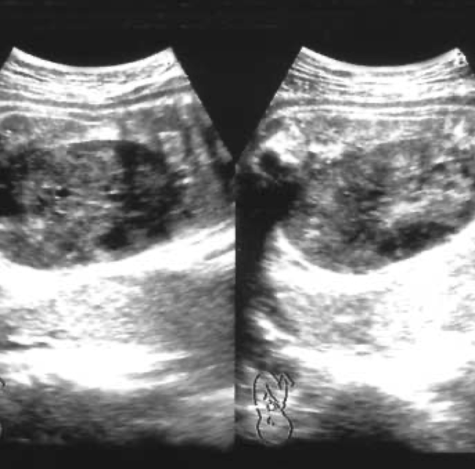
A 31-year-old woman was transferred to our hospital at 34 weeks of gestation due to sonographic picture of abdominal mass in her fetus associated with oligohydramnios. A previous C.S. had been carried out for a breech presentation. Her routine tests, including CBC, viral markers, urinalysis and $TORCH screen, and others, yielded nonspecific results, and the triple test indicated a low risk. At 26 weeks of gestation, ultrasonography revealed a fetal weight of 730 g, consistent with menstrual dates, with normal appearing fetus and sufficient amniotic fluid. Subsequently, the patient did not receive antenatal care for two months. Upon returning to the local clinic at 33 weeks of gestation, a fetal abdominal mass and reduced amniotic fluid levels were observed. The fetus was suspected of IUGR (1,370 g, below 10% for 33 weeks of gestation), prompting referral to our hospital. Upon admission, her vital signs were normal. Laboratory findings were unremarkable, and an indicated sonography was conducted and revealed a right renal large encapsulated abdominal mass which composed of multiple cystic and solid components. The other organs including the left kidney were normal. Following the monitoring of the fetal heart rate, recurrent and pronounced variable decelerations were observed, leading to the decision to conduct an emergency C.S.
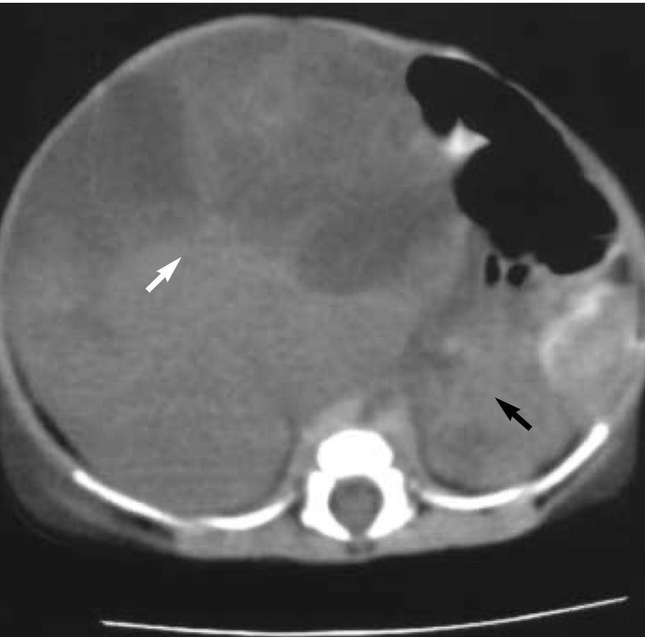
Upon admission to the NICU, the neonate exhibited a respiratiory rate 62/min, and blood pressure 69/43 mmHg. A physical examination revealed a prominent abdominal mass, measuring 7.5 cm in diameter on the right side of the abdomen, nearly occupying the abdominal cavity. The infant also presented with clubbed feet. Ultrasonographic examination and an abdominal CT scan unveiled a well-defined, heterogeneous, non-enhanced mass measuring 6.8 cm in diameter, originating from the right kidney, displaying both necrotic and cystic components. The left kidney appeared normal.
Within 24 hours of birth, the infant experienced anuria, severe anemia, hypoglycemia, and hypoalbuminemia. Both PT and aPTT were prolonged. Despite rigorous treatment, the infant failed to produce urine, and generalized and pulmonary edema ensued. On the second day, gastrointestinal hemorrhage and hypotension developed, leading to resuscitation and intubation. Due to the simultaneous presence of multiple severe systemic conditions, radical nephrectomy could not be performed. Laboratory findings indicated a general deterioration, with renal failure setting in. Hyperkalemia, hypoglycemia, thrombocytopenia, anemia, and poor coagulation profiles (aPTT undetectably prolonged) persisted despite extensive therapeutic interventions.
Unfortunately, the infant succumbed at four days of age, succumbing to respiratory failure and DIC.


Case Answer (December 2023)
Congenital mesoblastic nephroma (CMN)
CMN is the most common renal tumor in infancy, accounting for 3-6% of all childhood renal masses and 50% of all solid tumors in the neonatal period. Current opinion favors the classification of mesoblastic nephroma as a distinct, and usually benign neoplasm, arising from the renal parenchyma.
Sonographically, mesoblastic nephroma may present as a large (4 to 8 cm), unilateral renal mass with nodular densities, or as diffuse renal enlargement. These tumors are predominantly solid, but cystic areas are occasionally seen. Unlike Wilms’ tumor, there is no well-defined capsule, most likely due to hemorrhage with subsequent cystic degeneration.
Radical resection of the tumor is the treatment of choice, which is usually curative. However, a small percentage of patients with these tumors experience local recurrence or distant metastasis due to inadequate resection or cellular or atypical CMN, which is potentially a malignant tumor.
According to the literature, polyhydramnios was observed in almost 40% of the CMN cases, and acute fetal distress occurred in 25% of fetuses with CMN.
