Case Quiz (July 2022)
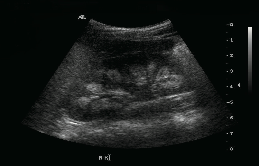
This patient was followed because of hypocalcemia, hypokalemia, and hypomagnesemia. The disorder was detected soon after birth. She had generalized seizures on the 14th day after birth. Her initial laboratory results showed hypocalcemia, hypomagnesemia, and a low level of PTH. Oral calcium, magnesium, and vitamin D supplements were prescribed and the seizures were controlled. However, if she stopped her medication, she had a seizure. Thiazide was added because of hypercalciuria and medullary nephrocalcinosis (Figure)
At 13 years of age, she suffered weight loss from 38 kg to 33 kg and a loss of appetite. After 4 days of nausea and vomiting, she attended hospital. Laboratory testing showed a serum calcium level of 15.9 mg/dL and serum phosphorus of 3.9 mg/dL. Her serum creatinine was 1.4 mg/dL. She had been prescribed calcium carbonate and alfacalcidol before admission. Medication was discontinued until serum calcium and phosphorus levels were below the normal range, after which the medications were restarted.
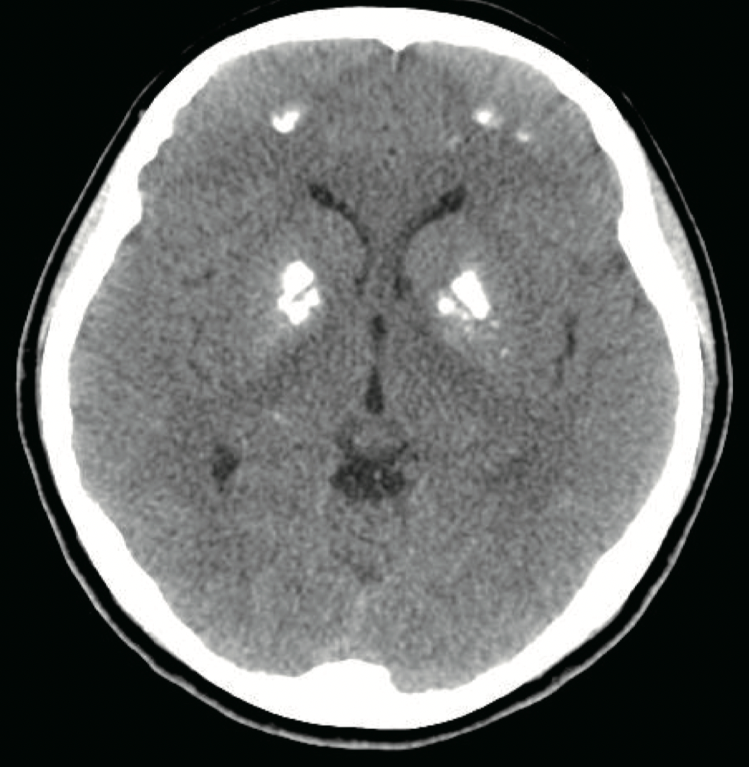
At 15 years old, the patient visited hospital complaining of dizziness, nausea, and vomiting. Her serum calcium level was 18.4 mg/dL and her serum potassium 2.8 mg/dL. Calcification of the subcortical and basal ganglia of both frontal lobes was found on brain computed tomography (Figure). Her medication at admission was calcium acetate, alfacalcidol, hydrochlorothiazide, and amiloride. A month before the second hypercalcemic episode and the dose of alfacalcidol had been increased because her serum 25-hydroxy-vitamin D3 (25-OHVitD3) concentration was just above the lower limit of the normal range. All medication was discontinued and her serum calcium and potassium concentrations gradually normalized, but because of persistent dizziness she was transferred to our hospital. On the day after transfer, she complained of paresthesia of her hand and her serum calcium concentration was 8.2 mg/dL. Alfacalcidol and calcium carbonate were administered and the dose of medication was slowly increased . Alfacalcidol was changed to calcitriol and she was discharged.
After informed consent was obtained, whole genome sequence analysis was done


Case Answer (July 2022)
Autosomal dominant hypocalcemia (ADH) with Bartter Syndrome (BS) is due to activating mutations in the CaSR can cause BS.
BS is a genetically heterogeneous disorder with a unifying pathophysiology of decreased salt absorption caused by different mutations throught the different renal tubules. The clinical presentations and onset timing of Bartter’s phenotype differ according to the type of mutation.
In this patient, hypokalemia occurred at 13 years old and was corrected with small doses of oral potassium. Patients with A843E or C131W mutations show the phenotype of BS including nephrocalcinosis, hypokalemia, hypomagnesemia, metabolic alkalosis, hyperreninemia, and hyperaldosteronemia as well as hypocalcemia. The characteristics of BS were expressed mildly in this patient. Serum magnesium concentrations were in the subnormal to lownormal range. Serum potassium concentrations maintained in the normal range until hypokalemia appeared.
This patient had three hypercalcemic episodes. Hypercalcemic episodes during the treatment of ADH have been reported previously, and occurred in patients who had taken a high dose of alfacalcidol. During her first hypercalcemic episode, our patient suffered fever and severe weight loss. The volume loss and the continuous medication might have caused the hypercalcemia. Before the second event, her dose of alfacalcidol was increased because of low serum 25-OHVitD3 , although her serum calcium concentration remained in the normal range. Thus, an overdose of alfacalcidol might have induced the hypercalcemia.
