Case Quiz (June 2023)
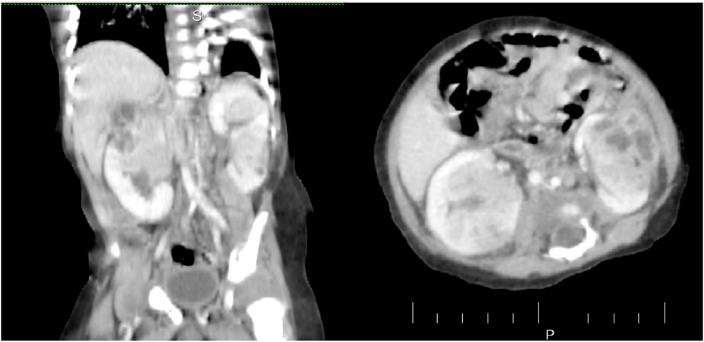
We received a referral from a pediatrician regarding a 3-month-old baby who had an incidental discovery of masses in both kidneys during an ultrasound examination. A follow-up CT scan without contrast indicated the presence of nephroblastoma/Wilms tumor. The infant had experienced a fever two weeks prior, for which the local general practitioner provided partial treatment with antibiotics. Currently, the baby is not running a fever and is fussy but able to consume food orally. There is no family history of cancer, and apart from a palpable non-tender mass in the left kidney, the physical examination did not reveal any noteworthy findings. Further investigations were conducted, revealing a total leukocyte count of 18,600 with 51% neutrophils, a hemoglobin level of 10.8, and a C-reactive protein level of 17.4. Other tests yielded normal results, including negative findings for growth in blood and urine cultures. Subsequently, a CT scan with contrast was performed, confirming the presence of nephroblastoma/Wilms tumor as the differential diagnosis (Figure).
Considering the potential diagnoses and the rarity of Wilm’s tumor at this age, an ultrasound-guided needle biopsy was performed on the renal mass.

Case Answer (June 2023)
Acute lobar nephronia
Acute lobar nephronia (ALN), also termed as acute focal nephritis or acute focal bacterial nephritis, is a non-liquefactive localized severe infection of the interstitium of the parenchyma of the kidney involving one or more of the renal lobes.
The clinical presentations of ALN include fever, flank pain, leukocytosis, pyuria, and bacteriuria, in most of the cases, which are similar to those with renal abscess or acute pyelonephritis. The blood parameters are similar to that of any systemic infections with neutrophilic leukocytosis and high.
Computed tomography (CT) is considered to be the most sensitive and accurate imaging modality for the diagnosis of ALN. CT scan with IV contrast has got better anatomical delineation providing more information on the lesion aspects, which goes in favor of ALN. The lesion presents as focal or global enlargement of the kidney. The corticomedullary differentiation is essentially blunted by edema with calyceal effacement.
