Case Quiz (May 2017)
A 2.5-year-old boy presented with a 3-day history of fever, anorexia, and emesis. Two days prior to admission, he developed nonpitting edema of his face, hands, and feet. On the day prior to admission, he developed a truncal rash and cracked lips. He was seen by his physician and referred for admission. There was no history of medication use.
Initial physical examination revealed an ill, irritable child. Temperature was 39.3 C, pulse 160 beats/min, blood pressure 118/70 mmHg, and weight 16.7 kg. Pertinent findings included: bilateral reddened conjunctivae, a "strawberry tongue", dry oral mucosa, clear pharynx, and an enlarged left cervical node. Heart, lung, and abdomen examinations were normal. The perineum was excoriated with an enlarged left inguinal node. There was nonpitting edema of the hands, feet, and face, and erythema of the palms and soles. The entire body was covered with an erythematous macular rash.
Laboratory evaluation showed: normal electrolytes, BUN 22 mg/dl, creatinine 1.0 mg/dl, AST 60 units/l, ALT 125 units/l, and total bilirubin 7.1 mg/dl. The white blood cell count was 26.8x 10³/mm3. The erythrocyte sedimentation rate was 76 mm/h, hemoglobin 11.7 g/dl, and platelets 438 x 103/mm3. An electrocardiogram was normal. An echocardiogram showed a prominent left coronary artery with normal cardiac function.
On hospital day 4, the patient became afebrile and urine output decreased to 1 ml/kg per hour. On hospital day 5, body weight had increased by 3 kg and total body edema was noted. Blood pressure was normal. Laboratory examination showed: sodium 129 mEq/l, potassium 7.9 mEq/l, carbon dioxide 21.5 mEq/l, BUN 26 mg/dl, creatinine 3.5 mg/dl, albumin 2.5 g/dl, cholesterol 150 mg/dl, and total bilirubin 0.9 mg/dl . The white blood cell count was 19.9x 10³/mm3 with 44% neutrophils, 36% bands, 5% lymphocytes, 7% monocytes, 5% eosinophils, and 3% atypical lymphocytes. Hemoglobin was 9.8 g/dl; C3 was 90 mg/dl. Urinalysis was negative for blood, or glucose. Microscopic examination showed many white blood cells, white blood cell casts, and oval fat bodies. Urinary protein was 16 mg/dl and creatinine 16 mg/dl. The fractional excretion of sodium was 7%.
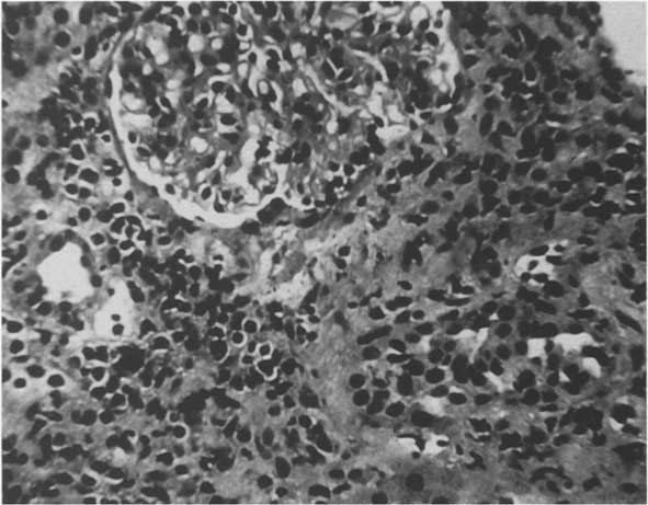
Abdominal ultrasound examination showed a prominent gall bladder and two large echogenic kidneys measuring 10.6 cm. There was no evidence of obstruction. A percutaneous renal biopsy was performed.
Case Answer (May 2017)
Kawasaki disease (KD) is an acute, multisystem vasculitis which affects predominantly young children below 5 years.This child was diagnosed as KD due to several clinical signs: fever, nonpurulent conjunctivitis, extremity changes, stomatitis, exanthems, lymph node enlarge-ment, hematological abnormalities, irritability, hepatitis, and cardiac findings.
Renal involvement in KD includes: elevations in blood urea nitrogen and creatinine concentrations, hematuria, proteinuria, and pyuria and occasionally echogenic, enlarged kidneys on ultrasound.
In our patient, acute interstitial nephritis occurred in the setting of KD. Our patient displayed anemia, acute renal insufficiency, oliguria, and tubular dysfunction. Urinalysis was significant for pyuria but not proteinuria, hematuria, or glucosuria.

