Case Quiz (May 2020)
A 12-year-old boy visited the hospital because of proteinuria and hematuria. The patient was diagnosed with ankylosing spondylitis (AS) at the same hospital 2 years prior. He was first administered NSAIDs and sulfasalazine, but the response was insufficient. His regimen was changed to etanercept and maintained for the past 1.5 years. No extra-articular manifestations of AS, such as uveitis, psoriasis or GIT involvement were reported. The patient had a history of asymptomatic renal stones. Intermittent microscopic hematuria was previously observed, but no additional testing was performed because it was considered indicative of asymptomatic renal stones. At the regular medical follow-up, microscopic hematuria and proteinuria were discovered, so he visited our hospital for a further evaluation.
On a physical examination, his vital signs were stable with a blood pressure of 140/80 mmHg, pulse of 80/min, respiratory rate of 20/min, and body temperature of 37℃. There were no other abnormal findings.
CBC revealed a WBCs of 6,300/㎕ (neutrophils, 71%; lymphocytes, 7%), Hb level of 13.5 g/㎗, and platelet count of 232,000/㎣. The following serum biochemistry test results were obtained: total protein, 7.4 g/㎗; albumin, 4.3 g/㎗; total bilirubin, 0.8 ㎎/㎗; AST, 36 IU/L; ALT 38 IU/L; and LDH, 412 IU/L. BUN level was 14 mg/dL and creatinine level was 1.02 ㎎/㎗. The erythrocyte sedimentation rate (ESR) was 10 ㎜/hr (normal, 0–16) and the high sensitivity C reactive protein (HS-CRP) level was normal. C3 level was 92.0 ㎎/㎗ , C4 level was 33.2 ㎎/㎗ and CH50 level was 48.1 ㎎/㎗ (normal, 23–46). The patient was positive for HLA-B27, while negative for rheumatoid factor, ANA, and ANCA. Furthermore, the patient was negative for hepatitis B antigen, hepatitis C antigen, and HIV, and was positive for hepatitis B antibody; venereal disease research laboratory tests yielded negative results.
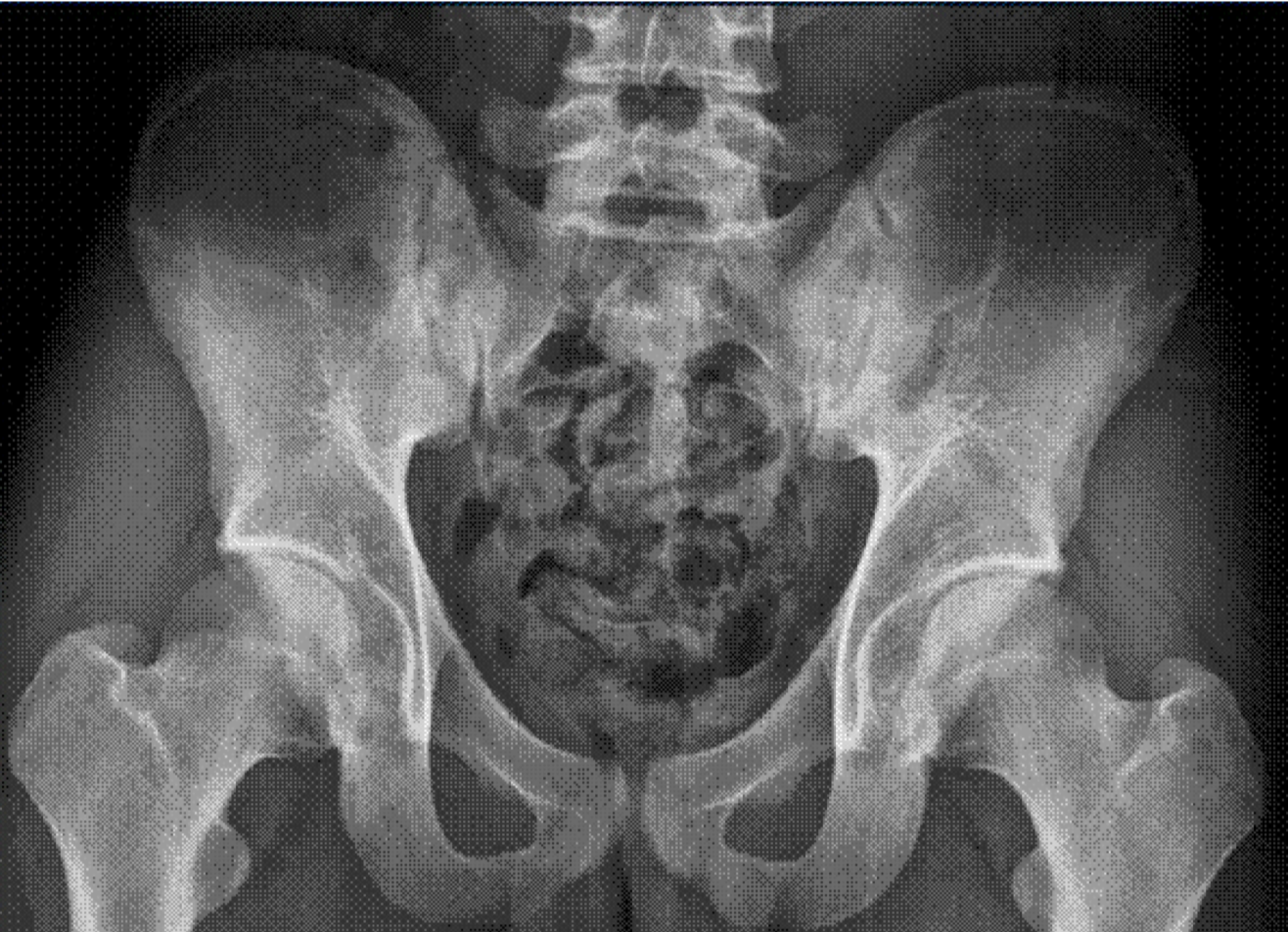
In urinalysis, the microscopic white blood cell count was 1–4 cells/high power field (HPF), many red blood cells and proteins were observed, and 24-hr urine protein level was 451 ㎎. The random urine protein/creatinine ratio was 287 ㎎/g. Mild bilateral sclerotic changes of the sacroiliac joint were observed on a pelvic radiograph.On renal U/S, the right kidney was 10.41 ㎝ and the left kidney was 11.07 ㎝, both within normal range, and no abnormal findings were observed in the renal parenchyma. However, a 0.9-㎝ calculus was observed in the Rt. kidney.
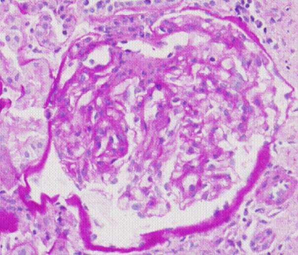
Renal biopsy was done
Case Answer (May 2020)
IgA nephropathy.In ankylosing spondylitis (AS), secondary renal amyloidosis is the most common form of kidney involvement (62%), followed by IgA nephropathy (30%) and mesangioproliferative glomerulonephritis (5%). Membranous nephropathy (1%), focal segmental glomerulosclerosis (1%), focal proliferative glomerulonephritis (1%), and treatment-induced nephrotoxicity also occur in rare instances.
Our patient had a well controlled disease and the proteinuria range does not coincide with secondary amyloidosis.
Renal biopsy conformed IgA nephropathy


